
Privates Investigator
- May 14, 2019
- 9 min read
Updated: May 21, 2019
Helping you become the detective who solves the mystery of what's going on "down there!"

Have you heard the abysmal statistic that roughly 44% of women polled were unable to identify the vagina on a diagram?! Whether this stat makes you laugh or cry, it should serve as a reminder that we have been slacking on our education about understanding our own genitalia. If this is already the case, it certainly makes sense that identifying what’s occurring in one’s vagina in the event of POP can lead to even more confusion.
We’re here to help!
Whether your hand mirror is your most cherished object, you refuse to look, or you’ve accidentally gone “Live” when your selfie cam was pointed south, we hope this blog helps you understand what you might be seeing!
Not only is it important to know your anatomy for your health and curiosity, but exposure to vulvas and vaginas that deviate from the textbook norm (which is often a hairless, not-impacted-by-childbirth-or-menopause vulva) can be key to improving genital body image.
So, ready to look at labia, visualize vulvas, and take a peek at POP?
Awesome! Let’s do this.
It should come as no surprise, given the nature of this blog, that we are about to feature pictures of vulvas with POP. It's certainly SFW for us but a NSFW warning seems appropriate before we continue!

We've been there.
Things feel... different. You're not exactly sure why or how, but something has changed. Maybe you decide to feel things out in the shower, or rummage through your medicine cabinet to find that hand mirror you haven't used in three years.
You notice that something has changed, but you realize that you lack a frame of reference: Did my vulva always look this way? Is this normal? What am I looking at? What am I supposed to be looking at?!!!
At this point, most of us head to Google and try to image search our way to clarity (spoiler alert: this rarely ends well!). Even with hundreds of images to scroll through, you're still not sure. What am I even looking at?!?!
Before we talk about what we're seeing when things have shifted, let's clarify the basic structures of the vulva and vagina:

Vulva is the term used to describe the external genitalia.
The vulval vestibule (or vulvar vestibule or vestibule of the vagina) is the part of the vulva between the labia minora into which the urethral meatus (not pictured here, the hole through which urine exists) and the vagina both open.
Labia majora are fattier and more external than the labia minora, the folds of skin between the labia majora. There's a ton of variation present in labia between people and this picture just shows one example, of course.
The clitoris is a sex organ that is the most erogenous zone in female bodies. It has internal and external components and, like us, it loves activewear - it comes with its own hood(ie)! The clitoral hood is formed by the labia minora.
The introitus is the opening to the vaginal canal. The hymen is a thin membrane that partially closes the vagina. In women whose hymens are no longer intact, hymenal remnants may remain, which often resemble small "tags" of skin. The hymen is relevant for prolapse assessment as it's the level by which descent is measured (POP grade is established based on the distance towards or beyond the level of the hymen).
The posterior fourchette is a fork-shaped fold of skin at the bottom of the entrance to the vagina that is designed to stretch.
The perineum is the muscular band and soft tissue between the vagina and the anus. It is frequently the site of trauma (tearing) during childbirth. Typically, in nulliparous women, the perineum appears slightly concave at rest, curving upward toward the ischial tuberosities (sit bones).
Not pictured:
Rugae are the series of ridges present in the walls the vagina. Those folds allow for the incredible expansion of the vagina. You'll feel them as ridged, bumpy tissue. Rugae are often absent or lessened when prolapse is present. The presence or absence of rugae is suggested to differentiate the specific site of the defect in POP. Sagging lateral vaginal sulci in the presence of anterior wall rugae suggest a paravaginal defect, or loss of lateral support. On the other hand, a central bulge with loss of vaginal rugae suggests a midline or central defect.
The urethra is a fibromuscular tube that conducts urine from the bladder to the exterior. In females, it's about 4 cm in length and 6mm in diameter, and fused with the anterior wall of the vagina. The hole through which urine passes through is called the urethral meatus.
GH + PB: It's possible you've heard the equation "gh+pb" but weren't quite sure to what that referred. The acronyms stand for "genital hiatus" and "perineal body," and they're measurements that are part of the POP-Q, a standardized assessment tool for quantifying POP. The genital hiatus (GH) is measured from the middle of the external urethral meatus to the posterior margin of the hymen (the part right before the perineum). The perineal body (PB) is measured from the posterior margin of the hymen to the mid‐anal opening. Increased gh measurements are associated with an increased risk of pelvic organ prolapse and levator muscle injury.
So, now that we're gotten that Health Education 101 module out of the way, let's talk POP!
Before we start, let's make sure we're all on the same page:
There is no currently no consensus on the definition of pelvic organ prolapse.
The International Urogynaecology Association (IUGA) and the International Continence Society (ICS) published a joint report in 2009 that described POP as
"the normal sensation, structure or function, experienced by the woman in reference to the position of her pelvic organs". This definition is quite vague, as are other definitions of POP! When women ask if their anatomy is "normal" (for someone who is postpartum, postmenopausal, etc.), it can be difficult to receive a straight answer. Beyond a certain age, virtually no one demonstrates "perfect" pelvic floor support and studies show that pelvic floor support beyond its anatomical origin (so, beyond stage 0 as defined by the POP-Q) is incredibly common in parous women, particularly those who have given birth vaginally.
Essentially, what qualifies as a "normal" postpartum vagina is up for debate and you'll receive ten different answers if you ask ten different providers. Some consider up to stage 2 POP (with the leading edge of the prolapse within 1cm above or below the hymen) "normal" for a postpartum person, while others are less inclined to characterize it as "normal". We're not going to spend the rest of the blog discussing normal (although you can read a brief IG post on our thoughts there!). Instead, we're simply going to describe the degree of descent.
Another thing to consider: Not all providers use a common nomenclature. Some refer to cystoceles and rectoceles while others use the terms anterior or posterior wall prolapse. Those who prefer to describe POP in relationship to the vaginal walls as opposed to specific organs (such as cystocele, referring to the bladder specifically) will note that we aren't always entirely sure what structure is behind the wall we're seeing. Meaning, what we perceive to be a rectocele (the rectum descended into the posterior wall) could in fact be the small bowel (referred to as an enterocele). What we're actually measuring is the wall itself, not the organ behind it.
Some will say that one's vaginal walls look "saggy" or "lax". Some have interpreted (and some providers have implied) that "vaginal laxity" or "looseness" is an entirely different entity from POP. The research on this suggests that "vaginal laxity" is associated with younger age, vaginal parity, symptoms of prolapse, prolapse bother and objective prolapse on POP-Q examination and imaging, suggesting that vaginal laxity may be considered a symptom of prolapse. The strongest associations were found with gh + pb and hiatal area on Valsalva maneuver, suggesting that vaginal laxity is a manifestation of levator ani hyperdistensibility."
So. What can we see from the outside?


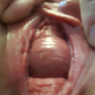
In this example. we can see the urethral meatus (where the teal star is) and a bulging of the anterior wall (where the purple star is). You can scroll the image to see it more clearly without the stars.
Also apparent is vaginal rugae (both on the visible bulge and on the lateral/posterior edge of the introitus/vagina. The vaginal rugae could be more prominent due to it being pushed out by the bulge behind it, as well be easier to see due to an enlarged introitus.
The descent of the anterior wall within 1cm of the level of the hymen would classify this as a grade 2 POP. This person is several years postpartum.

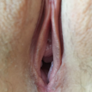
In this example, a protrusion of the anterior vaginal wall is demonstrated, within 1 cm of the hymen. A loss of rugae is evident on the anterior vaginal wall (at least the portion evident in this picture) with some rugae from the posterior wall "underneath".
This person was graded with a range of "no prolapse" from one provider to "grade 1-2" by another provider.


In these pictures, we can visualize both the anterior and the posterior wall prolapse.
In the first picture, the anterior wall POP seems more prominent, but the subsequent feature more of the posterior wall POP. As is visible in the third picture, when the anterior wall is retracted (sometimes done with a half speculum in clinical settings or with a finger), the posterior POP is more visible. The same would be true if the posterior wall were pushed back - we would be able to see more of the anterior wall.
The degree of descent observed by this person's provider was a grade 3-4 anterior wall POP and grade 3-4 posterior wall POP. Also potentially noteworthy is that this person is currently breastfeeding and less than a year postpartum.


The picture on the far left is taken in a supine position with a tilted/"lifted" pelvis (the note is from the owner of the vulva, and we assume it to mean either a slight decline position or a posterior tilting of the pelvis), the second is taken in a supine position with no pelvis shift and the third is taken standing. Particularly in the standing version, we can see some movement of the anterior wall. The person who supplied this picture noted her POP was evaluated as a "slight rectocele, minor cystocele and urethrocele." The cystocele/urethrocele are most evident in the standing photo on the right. (The person also noted a self-diagnosed urethral prolapse but that does not seem evident, at least in these pictures). What is helpful to note in these is how slight changes (from the left to the middle photo) can make a difference in the support of the pelvis and how it appears externally. Also, the impact of gravity (from left, which is supine, to right, which is standing) is demonstrated here, as well.


This example to the left shows another individual first in supine and then in standing. She reports that she is six months postpartum and has been diagnosed with a grade 2 cystocele.


In this example to the right, we can see the difference bearing down makes. In the right-hand picture, the anterior wall is more prominent. Note that the posterior rugae do not shift positions when the anterior wall does.
This presentation is a frequent topic of confusion, as the "walnut"-like appearance of the anterior wall does not resemble the more commonly identified cystocele images (smooth, round, water balloon). What we're seeing is descent at the level of the urethra (lower than bladder) with intact rugae, contributing to the ridged appearance.


This is another example of the difference bearing down can make. In the right photo, you can see movement of the perineum and are more clearly able to visualize the POP.
What accounts for the difference in POP presentation?

Several factors influence the appearance and presentation of one's POP.
The picture to the right illustrates how the pelvic floor musculature interacts with the urethra (the bladder would be above the tube), the vagina, and the rectum. Not pictured are the various connective tissue supports which also prevent the pelvic organs from descending into the vagina. You can visualize how the pelvic organs would have an opportunity to descend if any of these structures were damaged or weakened.
The appearance of one's POP is partially dependent on the specific site that has been compromised. The vaginal wall support system involves a complex interaction between muscular and connective tissue components. There are several levels of support and each one uniquely interacts with the architecture of the pelvis.
The degree of descent can be indicative of the extent of tissue damage or stretch. It can also be the case that the surrounding structures influence the visibility of the descent; for instance, two people might have the same degree of POP but the one belonging to the person with the larger introitus might be more visible externally.
Hormonal factors can influence the tissue's appearance and function, leading to changes in presentation of POP (less rugae, more irritation). One's unique anatomy (we did not all start with identical anatomy!) can further influence the appearance of POP.
Speaking of variety,
we think exposure to diverse genital imagery can serve educational purposes, as well as promoting positive genital body image. The following images were provided by people with varying degrees and types of pelvic organ prolapse. We are on a mission to provide people with POP (and people exploring whether they might have POP) with images that help them understand what they're looking at, and allow them to see themselves represented. Currently, a Google image search for POP typically includes only the most severe cases.
A variety of vulvas with various POP presentations:


We're working on a more expansive gallery and would love your help!
If you'd like to submit a photo, head to https://pop-up.filemail.com and it should automatically say it's sending to Haley Shevener (that's me!). For the "from" tab, go ahead and enter info@popuplifting.com (that keeps you anonymous). Then, in the subject and/or message, please state the type of POP and grade, attach your pic (cropped to include only your vulva - no other body parts) and hit send!
If you are interested in understanding your unique anatomy, a pelvic floor physical therapist is an excellent person to ask.
Often, they have mirrors available to walk you through the identification process. If you don't have access to in-person care, several virtual providers exist, including POP Up's own Annemarie Everett, PT, DPT, WCS. You can find her here: annemarieeverett.com


Comments